

 Since 2018
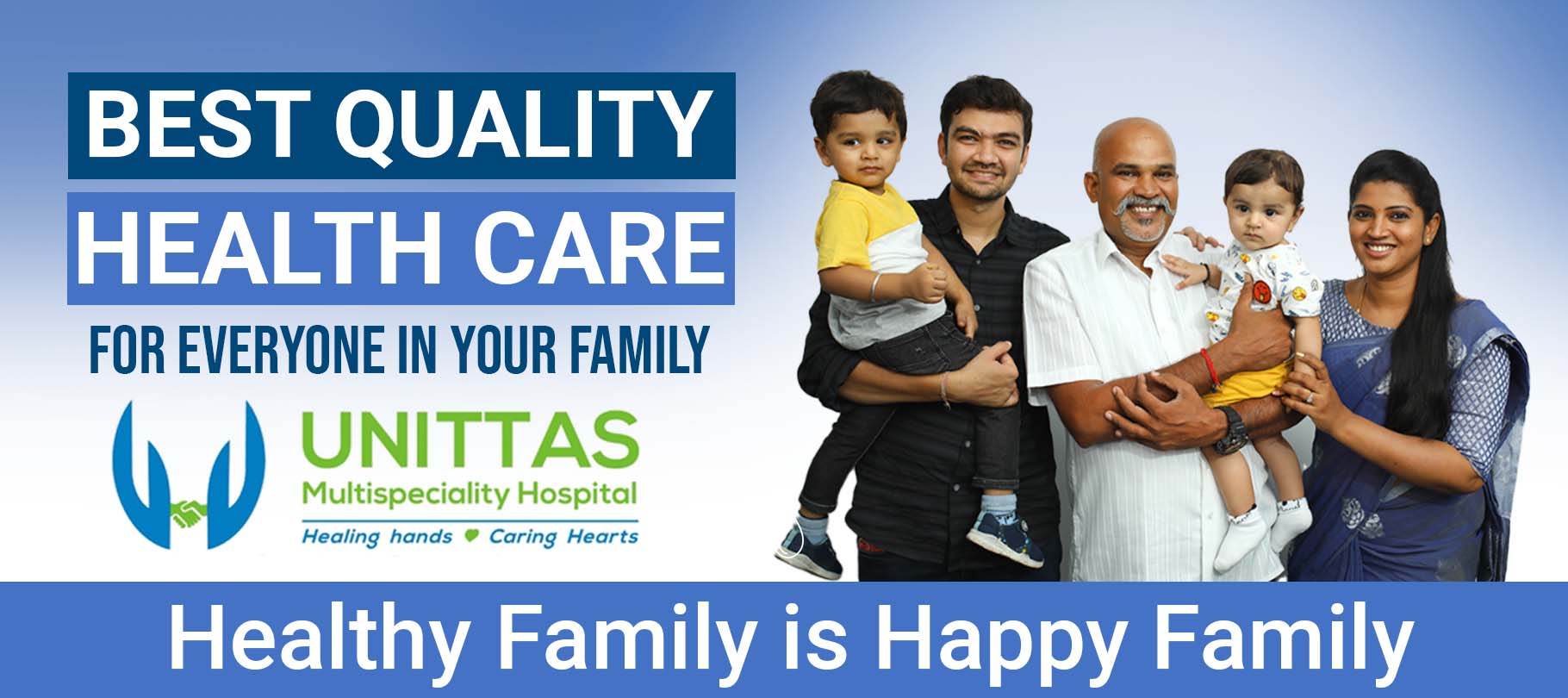
Multispeciality
24/7 Emergency
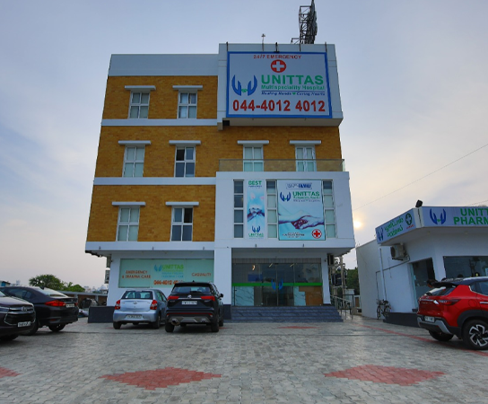
7, Duraiswamy Reddy St, West Tambaram, Chennai 600045
 Since 2018
7, Duraiswamy Reddy St, West Tambaram, Chennai 600045
 NABH
NABH
141/1, Kazhipattur Village, OMR Main Rd, Kelambakkam 603103
 Now Open
Indus Times Square, No 3/184, East Coast Rd, Uthandi 600119
Talk to our specialists from the comfort of your home. Secure video consultation, same-day slots available.
Video consult with lady doctors for PCOS, pregnancy, fertility, menopause & women's health concerns.
Consult Now Available NowExpert advice on UTI, kidney stones, prostate issues & male sexual health from experienced urologists.
Consult Now Available NowChild specialists for fever, feeding, vaccination, growth & newborn care — gentle, child-friendly.
Consult Now Available NowVideo consult for acidity, IBS, fatty liver, ulcers & digestive issues with expert gastro doctors.
Consult Now OMR
16y+ Exp
OMR
16y+ Exp
MBBS, MS (OG), DNB
Medical Director | Obstetrics & Gynaecology
 Tambaram
15y+ Exp
Tambaram
15y+ Exp
MS (OG), Diploma in Cosmetic Gynaecology
Cosmetic Gynaecologist
 ECR
2y+ Exp
ECR
2y+ Exp
MBBS, MRCOG
Obstetrics & Gynaecology
 Tambaram
15y+ Exp
Tambaram
15y+ Exp
MS (AIIMS), MCh (Urology)
Consultant Urologist
 OMR
12y+ Exp
OMR
12y+ Exp
MBBS, MS (Gen Surgery), MCh (Urology)
Urologist & Andrologist
 Tambaram
10y+ Exp
Tambaram
10y+ Exp
MBBS, MS, MCh (Urology)
Consultant Urologist
 Tambaram
20y+ Exp
Tambaram
20y+ Exp
MBBS, MS, MCh (SGE)
Surgical Gastroenterologist
 Tambaram
10y+ Exp
Tambaram
10y+ Exp
MBBS, MS, FIAGES, CCDM
General & Laparoscopic Surgeon
 Tambaram
10y+ Exp
Tambaram
10y+ Exp
MS, DNB, FMAS, FIAGES
General & Laparoscopic Surgeon
 Tambaram
8y+ Exp
Tambaram
8y+ Exp
MBBS, MS (General Surgery)
General & Breast Surgeon
Explore our most sought-after surgical treatments and procedures across all specialities.
 Hernia
 Inguinal Hernia
 Appendicitis
 Gallstone Removal
 Lipoma
 Breast Lump
 Axillary Breast
 Abscess
 Diagnostic Laparoscopy
 Kidney Stone
 Circumcision
 Varicocele
 Enlarged Prostate
 Hydrocele
 Bladder Stones
 Phimosis
 Vasectomy
 Testicular Torsion
 Ovarian Cyst
 Uterine Fibroids
 Endometriosis
 Hysterectomy
 Laparoscopic Hysterectomy
 Vaginal Hysterectomy
 Uterine Prolapse
 Uterus Removal
 Fibroid Embolization
 Diagnostic Hysteroscopy
 Rectocele
 ERCP
ERCP
 Gastric Bypass
 Gastric Band
 Sleeve Gastrectomy
 Adrenalectomy
 Rectal Polyps
 Knee Replacement
-injury-treatment-chennai.svg) PCL Injury
PCL Injury Carpal Tunnel
 Egg Freezing
 Embryo Transfer
 Semen Freezing
 Varicose Veins
 Deep Vein Thrombosis
 Thrombolysis
 Venous Thrombectomy
 Piles
Piles
 Fissure
 Fistula
 Pilonidal Sinus
 Dialysis
Very satisfied with the treatment and services. The medical team was professional and attentive.
My dad underwent fistula surgery. Doctors and nurses handled everything with great patience.
Visited for master health checkup. Naveena clearly explained and helped in every checkup.
My experience with Unittas been wonderful. I took my mom for master health checkup their guidance and support was awesome.
Excellent hospital with caring staff. Dr. Binu was very professional and explained everything clearly about my treatment options.
Had hernia surgery at Tambaram branch. Recovery was quick and the staff took excellent care during my stay.
Schedule an appointment with our expert doctors today.


